.")
.")


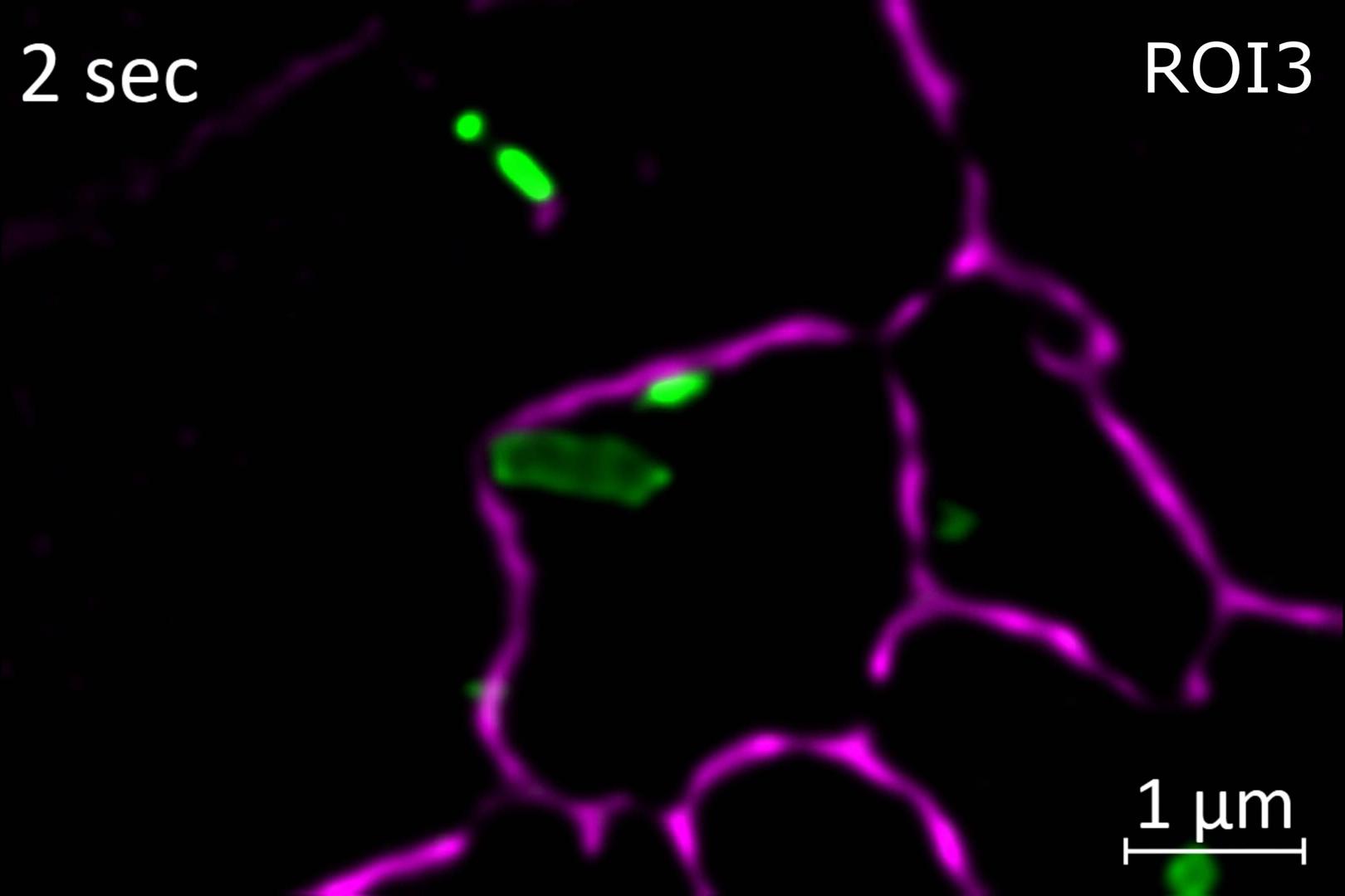
SIM² : doublez votre résolution SIM
SIM² est le nouvel algorithme révolutionnaire de reconstruction d'image qui augmente la résolution et la qualité du sectionnement optique des données de microscopie à éclairage structuré. SIM² est compatible avec tous les modes d'imagerie SIM de votre Elyra 7 et entièrement intégré au logiciel ZEN.
Contrairement aux algorithmes de reconstruction classiques, SIM² reconstruit l'image en deux étapes. Tout d'abord, une combinaison d'ordres, une réduction du bruit et un filtrage de suppression de fréquence sont effectués. Tous les effets résultant de ces manipulations d'images numériques sont traduits en une fonction d'étalement du point (PSF) numérique SIM. La déconvolution itérative subséquente utilise cette même PSF. Tout comme les avantages liés à l'utilisation de la PSF expérimentale pour la déconvolution des données de microscopie matérielle, l'algorithme SIM² est supérieur aux méthodes conventionnelles de reconstruction d'image en une seule étape pour la résolution, le sectionnement et la rigueur.


.")
.")
.")
.")


")
")


")
")


")
")


")
")