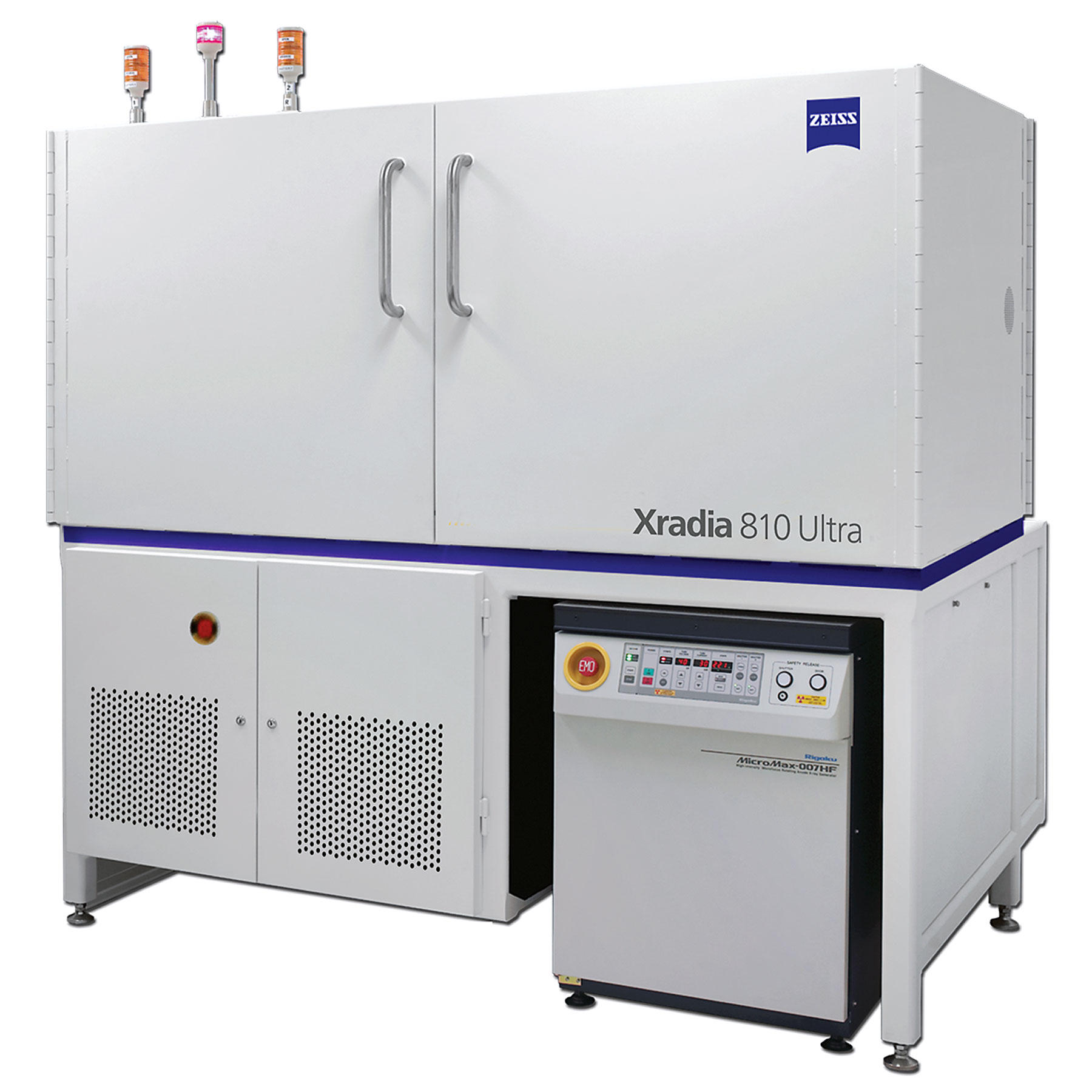
ZEISS Xradia Ultra
Imagerie par rayons X à l'échelle nanométrique : explorez à la vitesse de la science
Si la nanotomographie par rayons X synchrotron offre une imagerie 3D non destructive à l'échelle nanométrique, il convient d'appliquer un temps de faisceau très limité. Que diriez-vous de ne plus attendre le temps de synchrotron ? Imaginez disposer de capacités synchrotron dans votre laboratoire. Grâce à la série ZEISS Xradia Ultra, découvrez les microscopes à rayons X 3D non destructifs (XRM) qui offrent une résolution à l'échelle nanométrique avec une qualité comparable à celle d'un synchroton. Deux modèles sont disponibles : ZEISS Xradia 810 Ultra et ZEISS Xradia 800 Ultra, conçus pour offrir une qualité d'image optimale pour vos applications les plus fréquentes.


Optimisez vos recherches grâce à l'imagerie non destructive à l'échelle nanométrique
- Exploitez l'imagerie non destructive unique pour observer en 3D les phénomènes nanométriques dans leur environnement naturel.
- Bénéficiez du seul instrument qui comble le fossé entre les XRM à résolution submicronique (par ex. ZEISS Xradia Versa) et l'imagerie 3D à plus haute résolution, mais destructive, notamment les FIB-SEM.
- Utilisez des solutions in situ intégrées pour une imagerie par rayons X 3D / 4D non destructive de pointe dans votre laboratoire, avec une résolution réduite à 50 nm et une taille de voxel de 16 nm.
- Accélérez vos recherches en ajoutant ces capacités uniques à votre portefeuille analytique.


Un contraste et une image de qualité supérieure
- Observez les défauts en 3D sans détruire votre échantillon ni altérer les données avec des artefacts de tranchage.
- Grâce à l'absorption et le contraste de phase Zernike, révélez les détails avec la meilleure qualité de contraste et d'image possible. Combinez les données des deux modes pour révéler des caractéristiques inatteignables avec un seul contraste.
- Xradia 810 Ultra et Xradia 800 Ultra visent tous deux une qualité d'image optimale pour vos applications les plus fréquentes. La version qui vous convient le mieux dépend du matériau pour lequel vous souhaitez un contraste, un débit et une pénétration optimaux.
- Grâce à Xradia Ultra, bénéficiez d'une imagerie par rayons X à l'échelle nanométrique avec des capacités de type synchrotron.
Légende : tranche reconstituée en 2D d'une aiguille de pin en mode de contraste de phase Zernike (ZPC) (à gauche) et en contraste d'absorption (à droite).


Repoussez les limites de votre laboratoire
- Obtenez un nouveau niveau de compréhension grâce à des capacités de type synchrotron. Supprimez les obstacles à l'accès aux installations de synchrotrons. Obtenez des aperçus 3D équivalents à l'échelle nanométrique à des moments opportuns pour vous, dans votre laboratoire.
- Réalisez des études 4D et in situ jusqu'alors impossibles avec l'imagerie en laboratoire.
- Réalisez des essais mécaniques, thermiques, électrochimiques et environnementaux in situ.
- Utilisez des processus corrélatifs et connectez-vous à d'autres modalités (par ex. ZEISS Xradia Versa, ZEISS Crossbeam, analyses). Servez un large éventail d'utilisateurs d'installations d'imagerie grâce à une interface utilisateur rationalisée comprenant une API Python dédiée.
Légende : Structure de nanoréseau imprimée en 3D, imagée en contraste de phase Zernike avant les expériences de compression in situ. Avec l'aimable autorisation de : R. Schweiger, KIT, Allemagne (largeur d'échantillon 30 µm).


Illustration de la trajectoire du faisceau des microscopes à rayons X ZEISS Xradia Ultra.
Illustration de la trajectoire du faisceau des microscopes à rayons X ZEISS Xradia Ultra.
Profitez des avantages de l'architecture adaptée au synchrotron en utilisant :
- des condensateurs capillaires réfléchissants correspondant aux propriétés de la source pour obtenir une image à la densité de flux maximale ;
- des objectifs à plaque à zone de Fresnel qui, associés à des techniques de nanofabrication brevetées (US 8526575 B1 et US 9640291 B2), permettent d'obtenir des optiques présentant les meilleures résolution et efficacité de focalisation pour vos recherches ;
- un anneau de phase pour le contraste de phase Zernike afin de visualiser les détails dans des spécimens à faible absorption ;
- des détecteurs à haut contraste et à grande efficacité basés sur des scintillateurs, couplés optiquement à un détecteur CCD pour vous donner le meilleur signal dans votre temps expérimental limité ;
- la possibilité, au fur et à mesure de la rotation du spécimen, de collecter des images sur un éventail d'angles de projection, et de les reconstruire en un ensemble de données tomographiques 3D.


Applications
Découvrez comment réaliser l'imagerie d'échantillons provenant de domaines de recherche aussi variés que les sciences des matériaux, les sciences de la vie, les géosciences, etc.
Matériaux énergétiques


Matériaux d'ingénierie


Polymères et matériaux mous


Sciences de la vie


Électronique


Sciences de la terre


Accessoires


Résolution d'imagerie approximative pour les tests in situ, classement par épaisseur et transparence de l'échantillon. ZEISS Xradia Ultra comble le fossé entre la résolution nanométrique du MEB/TEM (limitée à l'imagerie de surface ou aux échantillons extrêmement fins) et la tomographie à l'échelle du micromètre.
Résolution d'imagerie approximative pour les tests in situ, classement par épaisseur et transparence de l'échantillon. ZEISS Xradia Ultra comble le fossé entre la résolution nanométrique du MEB/TEM (limitée à l'imagerie de surface ou aux échantillons extrêmement fins) et la tomographie à l'échelle du micromètre.
Expériences in situ à l'échelle nanométrique
Comblez le fossé des tests in situ
La recherche sur les matériaux étudie les propriétés qui apparaissent dans des conditions non ambiantes ou sous l'effet de stimuli externes. Si vous cherchez à observer les changements microstructuraux et à les relier aux performances du matériau, les méthodes d'essai in situ vous aident à atteindre cet objectif. Il est tout aussi important d'imager ces changements en direct et d'étudier des volumes d'échantillons représentatifs des propriétés en vrac.
Particulièrement adapté aux expériences in situ et à l'imagerie à l'échelle nanométrique, Xradia Ultra image des structures 3D de manière non destructive, en laboratoire, sur des échantillons de taille représentative des propriétés en vrac, mais dont la résolution correspond aux phénomènes à l'échelle nanométrique.


Observez vos spécimens in situ dans leur environnement naturel
Comprenez comment les événements de déformation et les défaillances sont liés aux caractéristiques locales à l'échelle nanométrique. En complétant les méthodes d'essais mécaniques existantes, obtenez des informations sur le comportement à plusieurs échelles de longueur. ZEISS Xradia Ultra Load Stage permet de réaliser des tests nanomécaniques in situ (compression, tension, indentation) uniques, en utilisant l'imagerie 3D non destructive. Étudiez ainsi l'évolution des structures intérieures en 3D, sous charge, jusqu'à une résolution de 50 nm.


Réalisez des expériences de chauffage in situ
Étudiez les changements de matériaux à l'échelle nanométrique tels que les processus de dégradation, l'expansion thermique et les transitions de phase à des températures élevées. La platine chauffante Norcada pour ZEISS Xradia Ultra assure une imagerie 3D non destructive à l'échelle nanométrique à des températures d'échantillon élevées. La technologie de chauffage MEMS chauffe l'échantillon dans l'air jusqu'à 500 °C. Sa conception flexible chauffe ou polarise l'échantillon avec la même unité.


Profitez du LaserFIB pour préparer les échantillons rapidement et en toute simplicité
Même si vos régions d'intérêt (ROI) sont profondément enfouies, accédez-y rapidement ou produisez facilement des échantillons en forme de piliers pour effectuer des tests avec ZEISS Xradia Ultra ou au synchrotron. Utilisez le LaserFIB qui combine un FIB-SEM ZEISS Crossbeam avec un laser femtoseconde (fs) à impulsions ultra-brèves, pour des processus corrélatifs sur plusieurs échelles de longueur. Trouvez vos ROI, par exemple à partir d'ensembles de données de microscopie à rayons X 3D précédemment acquis, et ciblez-les pour une analyse plus approfondie à l'aide du processus Cut-to-ROI. Utilisez le laser fs pour découper des millimètres de matériau et produire des échantillons à analyser avec Xradia Ultra. Ensuite, exploitez les capacités du FIB-SEM pour le fraisage à l'échelle nanométrique et micrométrique, la tomographie, l'imagerie et les analyses avancées.


Image de pile à combustible à oxyde solide réalisée sur Xradia Ultra.
Logiciel de visualisation et d'analyse : ZEISS recommande Dragonfly Pro
Cette solution logicielle d'analyse et de visualisation avancée fonctionne avec vos données 3D acquises avec différentes technologies, notamment les rayons X, le FIB-SEM et le MEB. Disponible exclusivement chez ZEISS, ORS Dragonfly Pro offre une boîte à outils intuitive, complète et personnalisable pour la visualisation et l'analyse de larges volumes de données 3D en niveaux de gris. Dragonfly Pro permet de naviguer, d'annoter, de créer des fichiers médias et de produire des vidéos à partir de vos données 3D. Effectuez un traitement d'image, une segmentation et une analyse d'objet pour quantifier vos résultats.


Paramétrez. Chargez. Explorez, scannez. Exécutez. C'est aussi simple que cela. Laissez l'interface utilisateur graphique vous guider dans la création de votre processus sans effort.
Créez des processus efficaces grâce à un logiciel convivial
Boostez votre productivité avec le système de contrôle innovant ZEISS Scout-and-Scan™ :rationalisez la configuration des échantillons et des scans. L'interface utilisateur vous guide tout au long de la procédure d'alignement de l'échantillon, de recherche de régions d'intérêt et de configuration des scans 3D. Grâce aux recettes, configurez plusieurs scans du même échantillon pour imager diverses régions d'intérêt ou combiner différents modes d'imagerie. Ce système facile à utiliser est idéal pour un environnement de laboratoire central dont les utilisateurs présentent parfois des niveaux d'expérience très variés. Les utilisateurs avancés obtiennent un contrôle total du microscope pour des tâches d'imagerie personnalisées ou l'intégration dans des expériences in situ à l'aide d'une API Python intégrée.
Applications liées
Téléchargements
-
-


ZEISS Xradia Ultra Family
Nanoscale X-ray Imaging: Explore at the Speed of Science
Taille du fichier: 10 MB -

ZEISS ORS Dragonfly
Outstanding 3D visualization with best-in-class graphics
Taille du fichier: 689 KB -

ZEISS Xradia Ultra Family - Flyer
Nanoscale X-ray Imaging: Explore at the Speed of Science.
Taille du fichier: 816 KB
-
-
-

A Brief Comparison of Computed Laminography versus 3D X-ray Microscopy
for Electronics Failure Analysis
Taille du fichier: 1 MB -

In Situ Observation of Mechanical Testing
at the Nanoscale
Taille du fichier: 1 MB -

X-ray Nanotomography in the Laboratory
with ZEISS Xradia Ultra 3D X-ray Microscopes
Taille du fichier: 6 MB -

3D Drill Core Scout and Zoom
For Gold Mineralization Characterization
Taille du fichier: 1 MB -

3D X-ray Imaging in Life Science Research
An Introduction to Capturing the 3D Structure of Biological Specimens Using X-rays
Taille du fichier: 3 MB -

In situ 3D Imaging of Crack Growth in Dentin
at the Nanoscale
Taille du fichier: 1 MB -

In situ Uniaxial Compression
Of Single Crystals of HMX explosive during 3D XRM Imaging
Taille du fichier: 988 KB
-