ZEISS Xradia Context MicroCT La plateforme microCT la plus avancée du marché
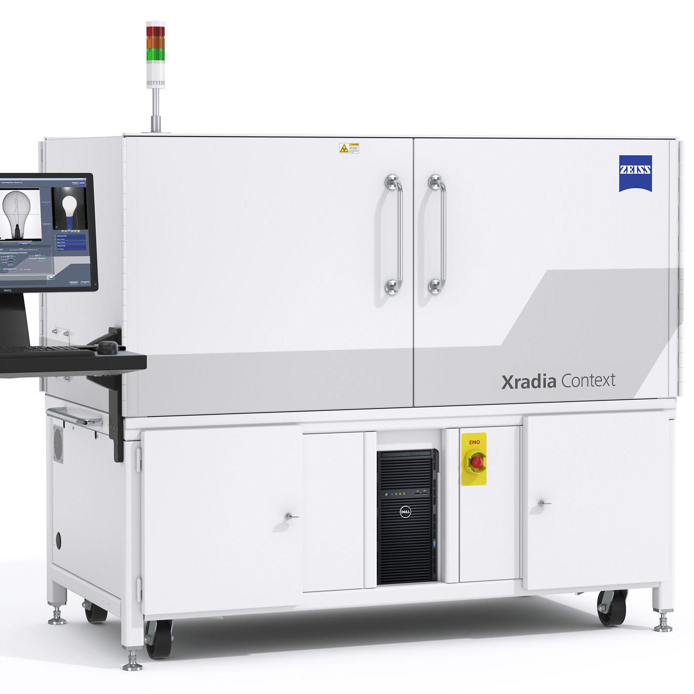
Xradia Context® de ZEISS est un système de tomographie par microcalcul (microCT) simple d'utilisation pour analyser tous types d'échantillons. Un détecteur de haute densité permet de visualiser en haute résolution les détails les plus fins, même dans des volumes d'imagerie relativement importants. Le système est doté d'un large champ d'observation, d'une fonction de montage et d'alignement rapide des échantillons et d'un processus d'acquisition simplifié. Il offre des temps d'exposition et de reconstruction des données rapides.


Imagerie 3D avec contexte global
- Xradia Context est synonyme de qualité d'image, stabilité et facilité d'utilisation. Il propose un environnement de processus performant, ainsi qu'un balayage à haut rendement.
- Un détecteur de haute densité de six mégapixels vous permet de résoudre les détails les plus fins dans leur contexte 3D complet, même lors de volumes d'imagerie relativement importants.
- Visualisez des structures enfouies de manière non destructive et en 3D pour l'analyse des processus, de construction et de défaillances.
- Maximisez l'agrandissement géométrique avec de petits échantillons pour identifier et caractériser des structures à l'échelle du micron avec un contraste et une clarté élevés.
- Bénéficiez d'un processus d'acquisition simplifié, d'un court temps d'exposition et d'une reconstruction des données rapides.
Vue en coupe virtuelle de l'intérieur d'un convertisseur catalytique intact.


Basé sur la plateforme réputée Xradia
- Xradia Context microCT repose sur des années de développement et une stabilité éprouvée, puisqu'il est construit sur la même plateforme que la gamme Xradia Versa.
- Découvrez un système ciblé sur les avancées en matière d'acquisition et de reconstruction des données en haute résolution et haute qualité.
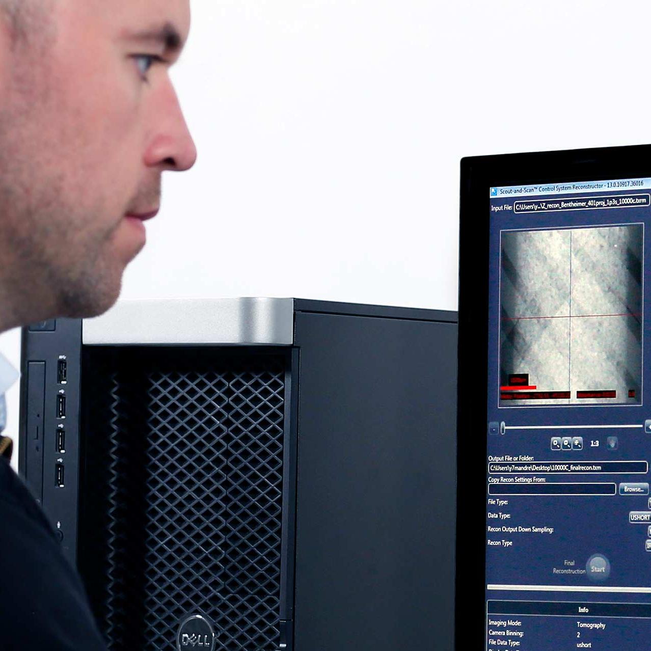
- Le système de contrôle convivial Scout-and-Scan vous fournit un environnement de processus efficace.
- Montez et alignez rapidement vos échantillons ou développez votre système avec Autoloader, disponible en option pour le traitement automatisé et la numérisation séquentielle de 14 échantillons.
- Le kit in situ permet des études en 4D et in situ en vue de mesurer les évolutions de la microstructure des matériaux dans des conditions variables.
- Ajoutez la Advanced Reconstruction Toolkit pour accélérer encore le rendement en conservant une excellente qualité d'image.
Scan Context microCT d'une montre intelligente intacte.


Convertible en microscope Xradia Versa (XRM)
- Votre instrument doit évoluer en fonction de vos besoins en imagerie. Xradia Context, au sein de la gamme de systèmes d'imagerie à rayons X ZEISS, bénéficie de l'engagement permanent de ZEISS à étendre les capacités et les fonctionnalités de ses systèmes sur le terrain.
- Ce concept ouvre la voie à une imagerie tomographique en 3D prête à évoluer en fonction de vos besoins : votre Xradia Context microCT est le seul microCT convertissable à tout moment en CrystalCT, en une plateforme 5XX, voire en microscope à rayons X 3D (XRM) ZEISS Xradia 620 Versa.
Imagerie du champ d'observation complet d'une mâchoire d'ours
Domaines d'application
-


Électronique
Scan Context microCT 3D d'une enceinte connectée. Visualisez de manière non destructive et en 3D des structures et des défauts enfouis pour des tailles d'échantillons variées, avec une résolution de premier ordre dans le champ d'observation choisi.
-


Fabrication
Rendu 3D d'une ampoule électrique. Context peut être utilisé pour effectuer un balayage complet des échantillons afin de vérifier leurs structures internes ou d'identifier des défauts dans les composants fabriqués.
-


Sciences de la vie
Vue en coupe d'un rendu 3D d'un embryon de souris dans de la paraffine. Les structures internes sont visibles à un contraste élevé. Échantillon avec l'aimable autorisation de l'hôpital général du Massachusetts.
-


Sciences de la terre
Coupe transversale virtuelle dans un échantillon de roche hétérogène révélant différentes phases et porosités.
-


Sciences des matériaux
Coupe virtuelle d'une batterie lithium-ion recyclée et désemballée, révélant des dommages dans les couches de collecteur de courant et cathodique. Échantillon avec l'aimable autorisation des professeurs M. U. Sauer et M. E. Figgemeier, ISEA, université technique de Rhénanie-Westphalie à Aix-la-Chapelle
-


Métaux
Rendu 3D des résultats d'un balayage rapide d'une aube de turbine pour évaluer la géométrie et inspecter les défauts internes ou les fissures.


Aperçu de la technologie
-

Xradia Context microCT repose sur la technologie éprouvée et réputée de la plateforme Xradia Versa.
Une qualité d'image basée sur une technologie éprouvée
Profitez d'un contraste et d'une clarté d'image de haute qualité pour différencier facilement les phases et pour la segmentation et la quantification en aval de vos données.
L'excellente qualité des données dépend de plusieurs facteurs, dont les caractéristiques de la source, l'ajustement de l'énergie des faisceaux, la géométrie et la sensibilité du détecteur, le contrôle de l'environnement, la stabilité des mouvements et les vibrations, l'étalonnage du système et la précision de reconstruction. Xradia Context microCT a été conçu sur la même plateforme que la gamme éprouvée de microscopes à rayons X Xradia Versa et a bénéficié des mêmes avancées qui ont permis à Xradia Versa de devenir un standard de performances dans l'imagerie 3D par rayons X en laboratoire.
- Filtres à rayons X supérieurs de correspondance des échantillons pour contrôler le durcissement du faisceau
- Modes avancés de correction automatique de la dérive
- Algorithmes avancés de réduction du durcissement du faisceau
- Algorithmes avancés propriétaires supplémentaires pour une qualité d'image optimale
-

Protégez facilement votre échantillon pour optimiser vos expériences
SmartShield est une solution qui protège votre échantillon et votre microscope. Ce système automatisé de prévention des collisions fonctionne avec le système de contrôle Scout-and-Scan. Il vous permet de naviguer dans Xradia Versa avec plus de confiance que jamais. D'un simple clic, SmartShield crée une couche protectrice numérique basée sur les dimensions de votre échantillon.
Grâce à SmartShield, vous profitez de :
- L'amélioration de l'efficacité de l'opérateur grâce à une configuration d'échantillons simplifiée
- Une expérience utilisateur améliorée pour les novices et pour les utilisateurs avancés
- La protection de vos échantillons et de votre investissement
- Une qualité de numérisation sans compromis
-

Mode champ large
Le mode champ large (WFM) peut être utilisé pour acquérir des images dans un champ d'observation latéral étendu. Le large champ d'observation latéral peut fournir un volume en 3D trois fois plus grand pour des échantillons de grande taille, ou une densité de voxel plus élevée pour un champ d'observation standard. Tous les systèmes Xradia Versa disposent du WFM lorsqu'ils sont équipés de l'objectif 0,4x. En combinaison avec l'assemblage vertical, le WFM permet d'acquérir les images d'échantillons plus volumineux avec une résolution exceptionnelle.


Accessoires
Ajoutez des accessoires à votre microscope et augmentez ses capacités


Boîte à outils avancée de reconstitution
Rendement supérieur et qualité d'image unique
Technologies de reconstruction assistée par IA (intelligence artificielle) pour vos systèmes ZEISS Xradia. Une compréhension approfondie des principes physiques des rayons X et de leurs applications vous permettra de relever de manière inédite et innovante certains des défis les plus ardus en matière d'imagerie.


Autoloader
Optimisez l'utilisation de votre instrument
Optimisez l'utilisation de votre instrument et réduisez les interventions de l'utilisateur grâce à ZEISS Autoloader, disponible en option. Réduisez la fréquence d'interaction avec l'utilisateur et augmentez la productivité en exécutant plusieurs tâches. Chargez jusqu'à 14 stations d'échantillons, c'est-à-dire jusqu'à 70 échantillons, placez-les en file d'attente et laissez votre instrument fonctionner toute la journée, ou hors des heures de service.


Kit d'interface in situ
Repoussez les limites de la science
Les plateformes ZEISS Xradia peuvent intégrer une grande variété d'équipements in situ, depuis les cellules de flux à haute pression aux platines de tension, de compression et thermiques, en passant par les conceptions personnalisées. Au-delà des trois dimensions de l'espace, profitez de la nature non destructive des examens aux rayons X pour étendre vos recherches grâce aux expériences 4D.


Batterie lithium-ion
Visualisation et analyse
ZEISS recommande Dragonfly Pro
ORS Dragonfly Pro est la solution logicielle d'analyse et de visualisation avancée pour vos données 3D acquises avec différentes technologies, notamment les rayons X, le FIB-SEM, le MEB et la microscopie à hélium ionisé. Disponible exclusivement chez ZEISS, ORS Dragonfly Pro propose une boîte à outils intuitive, complète et personnalisable pour visualiser et analyser de larges volumes de données 3D en niveaux de gris. À partir de vos données 3D, Dragonfly Pro permet la navigation, l'annotation ou encore la création de fichiers médias – y compris la production de vidéos. Effectuez un traitement d'image, une segmentation et une analyse d'objet pour quantifier vos résultats.
Applications liées
Téléchargements
-
-

ZEISS Xradia Context microCT
Your X-ray System for Today with Assurance for Tomorrow in English
Taille du fichier: 6 MB -

3D X-ray Microscope Field Conversion and Upgrade Options
Taille du fichier: 2 MB -

Flyer: ZEISS Xradia Context microCT
Advance your R&D with high resolution, non-destructive 3D imaging
Taille du fichier: 792 KB -

Flyer: ZEISS Xradia Context microCT
Increase your profitability and productivity with class-leading performance
Taille du fichier: 825 KB -

ZEISS Mineralogic 3D for Mining - Flyer
Your geometallurgy goals realized with maximum efficiency
Taille du fichier: 677 KB -

ZEISS PhaseEvolve
Reveal contrast that has never been seen before
Taille du fichier: 2 MB -

ZEISS Solutions for Semiconductor Development, Manufacturing, and Analysis
Accelerating Digital Transformation and Innovation for Semiconductor Electronics
Taille du fichier: 13 MB
-
-
-

Resolution of a 3D X-ray Microscope
Defining Meaningful Resolution Parameters
Taille du fichier: 932 KB -

3D X-ray Imaging in Life Science Research
An Introduction to Capturing the 3D Structure of Biological Specimens Using X-rays
Taille du fichier: 3 MB -

The building blocks of our solar system
Studying the Winchcombe meteorite
Taille du fichier: 3 MB
-